At the office of Tiger Family Dental, we prioritize precise diagnostics as the foundation of effective, predictable dental care. Cone-beam computed tomography (CBCT) gives our team a three-dimensional perspective that conventional X-rays simply cannot provide, helping us identify anatomy, assess treatment sites, and plan procedures with greater confidence.
Our practice uses advanced CBCT imaging to obtain crisp, targeted 3D scans while keeping radiation exposure low. These scans are an important tool for delivering safer, more efficient treatment across a range of dental disciplines, from restorative work to surgical planning.
CBCT creates volumetric images that show depth, spatial relationships, and fine anatomical detail. Unlike two-dimensional radiographs, a CBCT scan captures the jaw, teeth, airways, and supporting bone structures in true 3D, which makes it easier to recognize impacts, root orientations, and complex root canal anatomy.
Because the information is volumetric, clinicians can review thin cross-sections, panoramic views, and 3D reconstructions from a single scan. That flexibility helps reduce guesswork when diagnosing issues that are hidden or ambiguous on regular X-rays.
Importantly, modern CBCT units are optimized for dental applications — they focus on the area of interest and limit exposure to surrounding tissue. This targeted approach helps the care team obtain the images needed for a clinical decision while maintaining patient safety.
When planning dental implants or surgical procedures, accurate measurement and spatial awareness are essential. CBCT lets us measure bone height and width, evaluate bone density, and visualize critical anatomical structures such as the inferior alveolar nerve and sinus cavities in relation to proposed implant sites.
These insights allow for more predictable implant placement and can reduce the need for exploratory steps during surgery. The scan data can also be used to design surgical guides or to inform guided implant workflows, which further enhances precision and efficiency.
Beyond implants, CBCT supports other surgical decisions — for example, assessing impacted teeth, evaluating cysts or other pathologies, and mapping complex anatomy prior to a procedure. With comprehensive 3D imaging, the clinical team can design safer, more conservative treatment approaches tailored to each patient.
CBCT is valuable across many specialties. In endodontics, it can reveal extra canals, root fractures, or periapical lesions that are difficult to see on standard films. For orthodontic planning, three-dimensional images help evaluate tooth positions, airway size, and jaw relationships — information that supports precise treatment planning.
Temporomandibular joint (TMJ) evaluations also benefits from CBCT, which can show bony changes, joint surface relationships, and condylar position with clarity. Similarly, airway assessments using CBCT help identify anatomical contributors to breathing concerns that may affect sleep or oral health.
Because CBCT supplies a single scan with multiple viewing options, it reduces the need for repeated exposures while providing a richer set of diagnostic data for varied clinical scenarios.
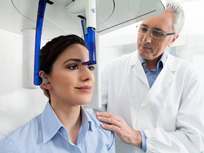
A typical dental CBCT appointment is brief and noninvasive. Patients sit or stand while the scanner rotates around the head, and a full-volume scan often takes less than a minute. The process is comfortable and requires no special preparation for most patients.
Modern CBCT systems are engineered to limit radiation dose by capturing only the region necessary for diagnosis. Our team follows established safety protocols, selects appropriate field-of-view sizes, and applies the principle of ALARA (as low as reasonably achievable) so every scan is justified and optimized for each patient’s needs.
Staff members operating the unit are trained in positioning and exposure techniques to ensure high-quality images with the fewest possible repeat scans. We also review each image carefully to confirm it meets diagnostic standards before concluding the appointment.
Acquiring a high-quality CBCT scan is just the first step. Interpreting the data requires clinical experience and a methodical approach. Our team examines the scan in multiple planes, correlates findings with clinical exams and other diagnostics, and uses the combined information to form a clear treatment plan.
CBCT data integrates smoothly into digital workflows, allowing for digital treatment planning, fabrication of surgical guides, and collaboration with specialists when complex cases require multidisciplinary input. The result is coordinated care built on objective imaging and shared clinical judgment.
Although CBCT is a powerful diagnostic tool, it is used selectively — not every situation requires a 3D scan. We evaluate each patient’s history and clinical presentation to determine when CBCT will meaningfully improve diagnosis or treatment outcomes.
In summary, cone-beam computed tomography enhances our ability to diagnose, plan, and deliver dental care with greater precision and confidence. If you’d like to learn more about how CBCT may play a role in your treatment, please contact our team for more information.


Cone-beam computed tomography (CBCT) is a three-dimensional imaging technology that captures volumetric data of the teeth, jaws, airway and surrounding bone. Unlike traditional two-dimensional radiographs, CBCT provides spatial relationships and depth information that reveal anatomy from multiple angles. This 3D perspective improves visualization of complex structures that are often obscured on standard films.
CBCT units used in dentistry are designed to focus on the region of interest and produce thin cross-sections, panoramic reconstructions and 3D renderings from a single scan. Because the data are volumetric, clinicians can reformat images in any plane to evaluate root morphology, bone contours and anatomical landmarks with greater confidence. The result is a richer diagnostic dataset that supports more precise treatment planning across specialties.
A CBCT scan is recommended when three-dimensional detail will materially affect diagnosis or the treatment plan, such as in implant planning, assessment of impacted teeth, complex endodontic cases, or evaluation of pathology. It is also useful when conventional X-rays are inconclusive or when accurate measurement of bone dimensions and anatomical relationships is required. The decision to use CBCT is clinical and based on whether the additional information will change care.
Using CBCT selectively helps avoid unnecessary imaging while providing crucial data when indicated. By targeting the field of view to the area of concern, clinicians obtain the images needed for safe, predictable care without exposing patients to more imaging than necessary. This selective approach aligns imaging choices with clinical benefit.
CBCT involves ionizing radiation, but modern dental CBCT systems are optimized to minimize dose by focusing the X-ray beam on a limited field of view and using efficient detectors. Clinicians apply the ALARA principle — as low as reasonably achievable — to choose the smallest field and lowest exposure settings that still provide diagnostic-quality images. Scans are used only when the expected diagnostic benefit outweighs the minimal radiation risk.
Operators are trained to position patients correctly and select appropriate protocols to reduce repeats and unnecessary exposure. For vulnerable populations such as children or pregnant patients, alternatives or modified protocols are considered and discussed with the patient to ensure safe imaging practices. The combination of contemporary equipment and careful protocol selection keeps doses low while preserving diagnostic value.
Preparation for a dental CBCT scan is minimal in most cases; patients are typically asked to remove eyeglasses, jewelry, removable dental appliances and hair accessories that could cause artifacts. Wearing comfortable clothing and arriving with any recent dental records or previous images can help the clinician correlate findings. There is generally no fasting or special medication required for the scan itself.
If a patient has specific mobility or positioning needs, informing the team before the appointment allows staff to arrange accommodations and streamline the process. Pregnant patients should notify the practice so the clinician can evaluate whether the scan is necessary or if alternative imaging is preferable. Clear communication before the appointment ensures a safe, efficient experience.
During a CBCT appointment the patient will sit or stand in the scanner with the head stabilized while the unit rotates around the head to capture a volumetric dataset. The scan itself typically takes less than a minute, although total appointment time may be longer due to positioning and brief image review. The procedure is noninvasive and generally well tolerated, with no injections or contrast agents needed for routine dental applications.
Technicians verify patient alignment and select the appropriate field of view and exposure parameters before initiating the scan to ensure diagnostic quality. After acquisition, the images are reconstructed and reviewed for clarity; if repeats are necessary they are kept to a minimum. The clinician then interprets the dataset in conjunction with the clinical exam to guide treatment decisions.
Reconstruction of a CBCT volume is usually available within minutes after acquisition, allowing the dental team to review the images during the same appointment or shortly thereafter. Clinicians examine the scans in multiple planes and correlate findings with the clinical exam to form a comprehensive treatment plan. This rapid access to high-resolution data supports timely decision-making for procedures such as implant placement or surgical referrals.
CBCT datasets can be exported to digital planning software to measure bone dimensions, create virtual implant placements, design surgical guides, or share with specialists for collaborative care. The images also serve as a baseline for monitoring anatomical changes over time. Integrating CBCT into the workflow improves precision and coordination across multidisciplinary cases.
CBCT greatly enhances implant planning by providing accurate measurements of bone height, width and density, and by showing the spatial relationship of critical structures such as the inferior alveolar nerve and sinus cavities. This information enables the clinician to select optimal implant size and angulation, avoid vital anatomy and determine whether bone augmentation is needed. Virtual planning based on CBCT data reduces uncertainty and supports more predictable surgical outcomes.
When appropriate, CBCT data can be used to fabricate surgical guides that translate virtual plans into precise clinical placement. Guided workflows reduce chairside guesswork, streamline procedures and can minimize surgical time and morbidity. By combining CBCT imaging with digital planning tools, clinicians achieve a higher level of accuracy in restorative-driven implant therapy.
In endodontics, CBCT can reveal complex root canal anatomy, accessory canals, root fractures and periapical pathology that may not be visible on two-dimensional images. The ability to view thin slices through a tooth and its surrounding bone helps diagnose causes of persistent symptoms and plan targeted retreatment or surgical intervention. This clarity reduces diagnostic uncertainty and supports more focused clinical strategies.
For temporomandibular joint (TMJ) assessment, CBCT provides detailed views of bony components such as condylar morphology, joint surface relationships and degenerative changes. While soft-tissue evaluation still relies on other modalities, CBCT is invaluable for assessing osseous pathology and guiding referral for multidisciplinary management. Combining CBCT findings with clinical examination yields a comprehensive picture of joint health.
CBCT has important limitations, including relatively poor soft-tissue contrast compared with medical CT or MRI, and susceptibility to metal artifacts from restorations or orthodontic appliances that can degrade image quality. Very large fields of view increase dose and may capture anatomy beyond the dental region, so clinicians choose the smallest practical field to address the clinical question. CBCT should not be used routinely for screening when less invasive imaging would suffice.
Clinical judgment determines appropriateness: if the expected 3D information will not change diagnosis or treatment, a two-dimensional radiograph may be more appropriate. Patients with certain medical conditions or those who are pregnant require individualized evaluation before proceeding. A careful benefit-risk assessment ensures CBCT is used when it provides clear diagnostic or therapeutic advantage.
CBCT interpretation is performed by trained dental clinicians who integrate the volumetric images with the patient history and clinical findings; in complex cases the practice may consult or refer images to specialists such as oral surgeons, endodontists or oral and maxillofacial radiologists. Interpretation requires experience with multiplanar review, recognition of normal variants and identification of pathology to ensure accurate clinical conclusions. When indicated, specialist collaboration enhances diagnostic accuracy and treatment planning.
Digital CBCT datasets are readily exportable in standard formats for secure sharing with consultants or laboratories, facilitating coordinated care. Patients are informed when images are sent to another provider and practitioners work together to review findings and agree on an appropriate treatment pathway. This collaborative model supports comprehensive, multidisciplinary care rooted in objective imaging data.
Looking to schedule your next dental visit or learn more about our services?
Getting in touch with Tiger Family Dental is simple! Our friendly team is ready to help you book appointments, answer questions about treatments, and address any concerns. Whether you’d like to call, or use our convenient online form, we’re here to assist you. Take the first step toward a healthy, confident smile — contact us today and experience the difference personalized dental care can make.