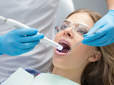
An intraoral camera is a compact, pen-sized imaging device designed to capture detailed color images from inside the mouth. Unlike traditional radiographs, intraoral cameras provide an immediate, full-color view of tooth surfaces, gum tissue, and restorative work. These live images are displayed on a chairside monitor so patients and clinicians can see the same view in real time.
This technology transforms a routine exam into a collaborative experience. By producing crisp, close-up photographs of specific teeth and soft tissues, the intraoral camera helps clinicians identify early wear, cracks, marginal decay around fillings, and areas of inflammation that might be hard to spot with the naked eye alone. It’s an important complement to other diagnostic tools, offering a surface-level perspective that X-rays and scans don’t always capture.
For patients, the value of intraoral imaging lies in visibility and clarity. What previously had to be described in professional terms can now be shown directly, making treatment needs easier to understand and discuss. In short, the intraoral camera brings transparency to the exam room and supports more informed decision-making.
During a dental exam, the intraoral camera is used as an extension of the dentist’s eyes. After a visual and tactile exam, the clinician will often use the camera to document suspicious areas or to record the condition of restorations and soft tissue. These images are captured quickly and saved to the patient’s digital record for comparison over time.
Images taken with the intraoral camera are used alongside digital X-rays and other diagnostic data to build a comprehensive picture of oral health. Because the photos are stored in the chart, clinicians can track small changes across visits—helping identify trends such as developing fractures, progressive wear, or gradual gum recession. This objective visual record strengthens preventive care and timely intervention.
The workflow is designed to be noninvasive and comfortable. The camera’s small head and gentle approach make it suitable for most patients, including those with limited jaw opening or sensitivity. In many cases, capturing a few targeted images early in the appointment saves time later by clarifying treatment priorities up front.
One of the most meaningful benefits of intraoral cameras is their role in patient education. Rather than relying on verbal descriptions, clinicians can point to high-resolution images to show exactly what they mean. This reduces confusion and helps patients visualize the scope and location of a problem, which improves comprehension and confidence in recommended care.
Images can be annotated or enlarged on-screen to highlight margins of restorations, hairline cracks, and plaque accumulation. This visual approach is particularly effective for explaining preventative measures: when patients see the precise areas where plaque builds up or where gum tissue is irritated, they are often more motivated to modify home care routines and follow clinical advice.
Because intraoral photographs can be shared with other members of the care team—such as specialists or hygienists—they help ensure everyone involved understands the situation in the same way. This consistency minimizes miscommunication and makes referrals and collaborative treatment planning more efficient and accurate.
From a clinical standpoint, intraoral cameras enhance diagnostic accuracy by revealing surface details that might otherwise be missed. Small fractures, leaking margins around restorations, and early carious lesions can be detected earlier when they are photographed regularly. Early detection frequently leads to less invasive treatment and better long-term outcomes.
Documentation is another strong advantage. High-quality images become a permanent part of the patient record, providing a visual timeline that supports clinical decisions. These images are valuable for monitoring the condition of restorations, evaluating tissue response after procedures, and confirming the healing progress following treatment.
When planning restorative or cosmetic work, intraoral photographs serve as a reference for shade selection, margin placement, and contouring. They are also helpful when communicating case details to dental labs or specialists, ensuring laboratory-fabricated restorations are matched and fitted with greater precision.
A typical intraoral camera exam is fast and comfortable. The clinician or hygienist will gently position the camera inside the mouth and capture several images of the teeth and gums. Because the device records color photographs rather than X-ray exposure, it is safe to use frequently as part of routine visits.
Patients can watch images appear on the monitor in real time, which often leads to immediate, clear explanations from the clinician. If any concerning details are found, the images remain accessible in the patient’s chart for future reference, comparison, and discussion at follow-up visits.
After imaging, the clinician will review the photos with the patient and outline recommended next steps, if any. The process is educational by design: patients leave the appointment with a clearer understanding of their oral health and actionable guidance tailored to their needs.
At Tiger Family Dental, we use intraoral cameras to make dental care more transparent, precise, and patient-focused. If you’d like to learn more about how intraoral imaging fits into our approach to exams and treatment planning, please contact us for more information.


An intraoral camera is a small, pen-sized imaging device that captures high-resolution color photographs from inside the mouth. It provides an immediate, magnified view of tooth surfaces, gum tissue, and existing restorations so clinicians and patients can see the same details in real time. Because it records surface-level images rather than relying on radiation, it complements other diagnostic tools by showing visual information X-rays do not capture.
Clinicians use intraoral cameras to document findings, educate patients, and monitor changes across visits. The images help identify early wear, cracks, marginal decay, and tissue inflammation that might be difficult to see with the naked eye. Overall, the technology supports clearer communication and more informed treatment decisions.
Intraoral photographs show surface color and texture while digital X-rays and CBCT scans reveal internal structures and bone relationships. Together these modalities create a fuller diagnostic picture: radiographs detect hidden decay and bone loss, CBCT provides three-dimensional anatomic detail, and intraoral images document visible surface conditions. This layered approach reduces diagnostic uncertainty and helps clinicians prioritize care based on both surface appearance and internal findings.
Using photos alongside radiographs also improves record keeping and case review. Visual images make it easier to compare surface changes over time, while radiographic data confirms underlying causes. The combination enhances accuracy in diagnosis, treatment planning, and interdisciplinary communication.
An intraoral camera excels at revealing subtle surface issues such as hairline cracks, marginal gaps around restorations, staining patterns, and early plaque accumulation. These surface signs often precede symptoms and can be important early indicators of structural weakness or developing decay. By capturing clear, magnified photos, clinicians can spot and document issues before they progress to more extensive problems.
The camera is also valuable for monitoring soft tissue conditions like localized inflammation, recession, and mucosal changes. Regular photographic records make it easier to detect gradual changes that are hard to notice during a single visual exam. Early detection frequently enables less invasive care and better long-term outcomes.
At Tiger Family Dental the intraoral camera is used as part of a comprehensive exam to supplement visual inspection and tactile assessment. After an initial check, the clinician or hygienist will capture targeted images of suspicious areas or restorations and store those photos in the patient’s digital record. This streamlined workflow is noninvasive and typically adds only a few minutes to the appointment while providing valuable documentation.
Those images are reviewed with the patient on a chairside monitor so the clinician can explain findings and discuss recommended next steps. Because the photos are saved, the team can compare them across visits to monitor trends and treatment response. This approach reinforces preventive care and helps patients make informed choices about their oral health.
Yes. Intraoral cameras are designed with small heads and gentle profiles to minimize discomfort and accommodate most mouths, including patients with limited jaw opening. The device records color photographs and does not expose patients to radiation, so it can be used frequently during routine visits without safety concerns. Capture times are short, making the process efficient and well tolerated by the majority of patients.
Clinicians take care to position the camera gently and explain each step so patients know what to expect. Because images appear on a monitor in real time, many patients find the experience engaging and reassuring rather than intrusive. The overall goal is to make imaging comfortable while delivering clearer information about oral health.
Visual images remove much of the ambiguity that can accompany verbal descriptions of dental problems by showing the exact area of concern. When clinicians enlarge or annotate photos on-screen, patients can see cracks, margins, plaque, and tissue changes clearly, which improves understanding and reduces confusion. This transparency helps patients weigh options with confidence and participate actively in care decisions.
Images also support better documentation of the discussion and recommended plan, so patients and clinicians share a consistent record of what was observed and why treatment is advised. Sharing photos with specialists or hygienists ensures everyone involved in a patient’s care interprets the situation the same way. The result is more coordinated, patient-centered treatment planning.
Intraoral images are saved directly to the patient’s digital chart where they become part of a longitudinal record. This makes it possible to track small changes—such as progressive wear, recurrent decay, or tissue recession—over months and years. Having a visual timeline supports preventive strategies and timely interventions that can limit the need for more extensive treatment later.
Stored photos are also useful when planning restorative or cosmetic procedures because they provide objective references for shade selection, margin placement, and contouring. Clinicians frequently use archived images to evaluate outcomes after treatment and to communicate detailed case information with dental laboratories or specialists. These records strengthen clinical decision making and quality control.
Yes. High-quality intraoral photographs help clinicians assess margins, contacts, surface morphology, and shade relationships before restorative or cosmetic work. These images provide visual guidance for selecting materials and designing preparations, and they can be shared with dental labs to improve the accuracy of fabricated restorations. For cosmetic cases, photographs document preoperative conditions and assist with shade matching and esthetic discussions.
During treatment planning, photos can be annotated to indicate margin placement, proposed contours, and areas requiring attention. This visual documentation improves communication among the clinical team, the patient, and laboratory technicians. As a result, restorative and cosmetic outcomes are often more predictable and closely aligned with patient expectations.
Patients can expect a quick, noninvasive process in which a clinician or hygienist gently positions the camera and captures several targeted images. The images appear on a chairside monitor in real time, allowing the clinician to explain findings immediately. Because the device records color photographs and not X-rays, there is no radiation exposure associated with the imaging itself.
After imaging, the clinician will review the photos with the patient and outline any recommended next steps or preventive measures. The images remain in the patient’s record for future comparison, which helps guide follow-up care. Many patients leave the appointment with a clearer understanding of their oral health and specific actions to take at home.
The frequency of intraoral photography depends on each patient’s risk profile and treatment needs, but many patients receive targeted photos during regular checkups or when a specific concern arises. Patients with a history of restorations, visible wear, orthodontic appliances, or periodontal issues often benefit from more frequent imaging so clinicians can monitor changes closely. Imaging can also be valuable before and after restorative or cosmetic procedures to document condition and outcomes.
Routine use is particularly helpful for patients who prefer visual feedback or who need objective documentation for long-term monitoring. Because images are easy to store and compare, they serve a wide range of clinical and educational purposes across preventive, restorative, and specialty care. When used thoughtfully, the technology supports better outcomes and more personalized care.
Looking to schedule your next dental visit or learn more about our services?
Getting in touch with Tiger Family Dental is simple! Our friendly team is ready to help you book appointments, answer questions about treatments, and address any concerns. Whether you’d like to call, or use our convenient online form, we’re here to assist you. Take the first step toward a healthy, confident smile — contact us today and experience the difference personalized dental care can make.